- Campbell Arnold
- Jun 17, 2025
- 6 min read

Updated: Jun 23, 2025

“Whole-body MRI & traditional single-cancer screening methods may synergistically enhance our ability to detect disease early.”
— Yosef Chodakiewitz, Medical Director at Prenuvo
Welcome to Radiology Access! your biweekly newsletter on the people, research, and technology transforming global imaging access.
In this issue, we cover:
Double the Contrast—Not the Dose
Low-Field MRI Gears Up for Big-League Neuroscience
Early Insights from Prenuvo's Whole-Body MRI for Cancer Screening Study
Positrigo raises $8.5M for ultra compact brain-only PET scanner
If you want to stay up-to-date with the latest in Radiology and AI, then don't forget to subscribe!
Double the Contrast—Not the Dose
How AiMIFY uses AI to enhance contrast in brain MRI without more gadolinium.

This week, I’m thrilled to highlight one of my own projects—just posted as a preprint on medRxiv! As many of you know, doubling gadolinium dose is known to improve lesion conspicuity, particularly for small or poorly enhancing lesions. However, higher doses have not become standard-of-care due to concerns over gadolinium retention, nephrotoxicity, and increased cost. Our new study presents a promising alternative: AiMIFY, a deep-learning-based algorithm that boosts contrast without requiring more gadolinium.
Developed by Subtle Medical in collaboration with Bracco Imaging, AiMIFY builds on prior work in contrast dose reduction and adapts it to amplify standard-of-care images. To validate the algorithm’s performance, we ran a retrospective, multi-center study across 110 patients with a variety of brain pathologies. Here are some key study details:
Standard Pre- and post-contrast scans were run through AiMIFY to generate enhanced versions.
Three board-certified neuroradiologists reviewed image sets, rating them on a variety of qualitative metrics.
We also quantitatively assessed contrast enhancement and used a physics-based method to quantify the contrast dose equivalence.
After our analysis, we found that:
AiMIFY significantly improved lesion conspicuity across all three quantitative metrics.
Had higher radiologist scores for lesion conspicuity, border delineation, internal morphology, and overall image quality.
Some readers found increased vessel conspicuity distracting, but any false positives were confidently ruled out using the standard dose images.
Contrast gains were approximately equivalent to a double-dose study, based on the physics-based analysis.
As first author Srivathsa Pasumarthi put it, “AiMIFY boosts the contrast signal, amplifying the existing signal in standard-of-care imaging.” And when you're searching for subtle lesions in cases like demyelinating disease or brain tumors, “identifying one new lesion can be a life changing outcome.” This study highlights how AI isn’t just a tool for cost savings or workflow optimization—it’s a way to get more from the images we already acquire.
Bottom line: AiMIFY uses AI to amplify contrast in brain MRI without increasing gadolinium dose—offering a safer, more effective way to boost lesion visibility when it matters most.
Low-Field MRI Gears Up for Big-League Neuroscience
How Hyperfine’s portable scanner could enable global-scale brain research.

Low-field MRI has long held promise for expanding access to clinical imaging beyond the hospital walls. Now, new research is showing it may also be ready for high-impact global neuroscience research. Two recent papers foreshadow how Hyperfine’s 64mT portable MRI system could enable large-scale neuroscience studies—one demonstrating the feasibility of diffusion tractography, and another outlining a robust quality control framework for a massive a multi-site, multi-continent research network.
In an arXiv preprint, a UK-led team showed that diffusion tractography—typically the domain of high-field MRI—can be performed on the 64mT Hyperfine system. Tractography allows researchers to map white matter pathways in the brain and plays a crucial role in studying development, neurodegeneration, and injury. Key findings from the study include:
Successful reconstruction of major white matter bundles in five healthy adults using a clinically viable scan time (<1 hour).
Strong correspondence between diffusion metrics derived from low-field and high-field scans.
The results validate the feasibility of tractography on portable scanners, opening up a significant new research tool on low-field devices.
Meanwhile, a second paper published in Human Brain Mapping by the UNITY project outlines a quality control framework for a 17-site, 12-country trial using Hyperfine scanners. Using CaliberMRI’s portable phantom, researchers validated image consistency across a range of environmental conditions—including differences in electromagnetic noise and temperature. Highlights from the QC study include:
Minimal geometric distortion across sites.
High scan success rates and consistent performance over time.
A scalable framework ensuring reproducibility in large international studies.
Together, these studies signal a new phase for portable MRI—not just as a tool for bedside diagnosis, but as an engine for global-scale neuroscience. By enabling advanced imaging in places far from high-end scanners, Hyperfine and similar technologies could help democratize access to research and reshape how and where we study the human brain.
Bottom line: New research shows low-field MRI can support advanced techniques like diffusion tractography and deliver consistent image quality across global multi-site studies.
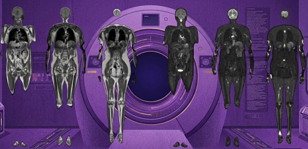
Early Insights from Prenuvo's Whole-Body MRI for Cancer Screening Study
Can they convince people to rethink head to toe cancer screening?

Could whole-body MRI become a powerful new tool for early cancer detection? Preliminary results from Prenuvo’s Polaris study—unveiled at the American Association for Cancer Research annual meeting—suggest it might. The retrospective analysis included 1,011 individuals who underwent non-contrast, whole-body MRI scans with diffusion-weighted imaging. Importantly, no participants had active cancer at the time of screening.
Based on direct-to-patient phone calls with a median 14 month follow-up, whole-body MRI led to 41 diagnostic tissue samples, with just over half confirming a new cancer diagnosis—yielding a 2.2% overall cancer detection rate. Other key findings include:
86% of detected cancers occurred in patients who reported no specific symptoms prior to their scan.
68% of cancers were located in areas not covered by standard screening guidelines.
Cancer detection increased with age, peaking at 3.0% in individuals aged 65–79. No cancers were found in those under 35 or over 79, but sample sizes were small in those groups.
Two false negatives (0.2%) were reported, both breast cancers, alongside 19 false positives that led to biopsy.
Beyond cancer, scans identified other clinically significant conditions—including aneurysms, benign masses, and liver or lung abnormalities—though only anecdotal evidence was provided.
While these early results are promising, real-world validation of whole MRI as a screening tool will require larger, prospective studies with standardized reporting frameworks and more complete follow-up data—a major limitation of the present work being the reliance on patient self-reports. Still, Prenuvo’s broader Hercules study—which plans to track 100,000 individuals over a decade—could be a major milestone in expanding early detection for cancers currently missed by standard screening tools.
Bottom line: Preliminary Prenuvo results suggest whole-body MRI can detect cancers not covered by screening guidelines, with a 2.2% overall detection rate.
Positrigo raises $8.5M for ultra compact brain-only PET scanner
Can they capture a segment of the growing dementia market?

Positrigo—an ETH Zurich spin‑off—is advancing functional brain imaging with its ultra-compact, brain-only PET scanner, NeuroLF. Following FDA and EU clearances, the company has closed an $8.5 million funding round led by HealthCap and Navivo. The company is gearing up to scale production and preparing for a commercial launch in North America and Europe.
Why a brain-only scanner? With amyloid and tau PET tracers gaining clinical relevance—and a new wave of Alzheimer’s therapies on the horizon—Positrigo is betting on dementia as its primary use case. The Alzheimer’s population is projected to reach 150 million globally by 2050, yet PET remains costly and infrastructure-heavy. NeuroLF offers a leaner, more affordable solution with minimal siting requirements, making it suitable for deployment in outpatient settings.
This investment marks an important step toward increasing access to functional brain imaging. With its compact footprint and patient-friendly design, NeuroLF could help bring advanced neuroimaging to memory clinics, community hospitals, and beyond—supporting earlier diagnosis and expanding access to care as demand continues to rise.
Bottom line: Positrigo’s $8.5M raise aims to bring compact, affordable brain PET imaging to the frontlines of dementia care in the US and EU.
Resource Highlight: RadPod
These days, who isn’t looking for work freedom and more money? A new teleradiology platform, RadPod, promises to be “the Uber-like, on-demand platform that liberates radiologists, giving them the freedom to work when they want, how they want, and where they want.” Radiologists can pick up reads at their convenience, with per-case 1099 pay and no long-term commitments.
Currently, RadPod seems to still be in an early rollout—serving eight hospitals in Illinois—but it’s a model worth watching as remote work and flexible staffing models continue to gain momentum. If you’ve tried RadPod or are considering signing up, I’d love to hear about your experience, please reach out!
Feedback
We’re eager to hear your thoughts as we continue to refine and improve RadAccess. Is there an article you expected to see but didn’t? Have suggestions for making the newsletter even better? Let us know! Reach out via email, LinkedIn, or X—we’d love to hear from you.
References
Pasumarthi, Srivathsa, et al. "Deep-Learning Based Contrast Boosting Improves Lesion Visualization and Image Quality: A Multi-Center Multi-Reader Study on Clinical Performance with Standard Contrast Enhanced MRI of Brain Tumors." medRxiv (2025): 2025-06.
Gholam, James, et al. "Diffusion Tensor MRI and Spherical-Deconvolution-Based Tractography on an Ultra-Low Field Portable MRI System." arXiv preprint arXiv:2506.04473 (2025).
Ljungberg, Emil, et al. "Characterization of portable ultra‐low field MRI scanners for multi‐center structural neuroimaging." Human Brain Mapping 46.8 (2025): e70217.
https://www.positrigo.com/positrigo-closes-financing-round-and-appoints-new-board-members/
Disclaimer: There are no paid sponsors of this content. The opinions expressed are solely those of the newsletter authors, and do not necessarily reflect those of referenced works or companies.