- Campbell Arnold
- Jul 15, 2025
- 4 min read


“Despite remarkable improvements in [MRI] over the last decades, CT continues to be the diagnostic instrument of choice for [fracture detection].”
— Fischer et al., European Radiology 2025
Welcome to Radiology Access! your biweekly newsletter on the people, research, and technology transforming global imaging access.
In this issue, we cover:
Philips Gains FDA Clearance for Image Acceleration Software
MRIguidance Aim to Bring Synthetic CT Into Routine Care
DIY MRI Workshop Returns in 2026—Free, Hands-On, and Open to All
If you want to stay up-to-date with the latest in Radiology and AI, then don't forget to subscribe!
Philips Gains FDA Clearance for Image Acceleration Software, SmartSpeed Precise
And what this means for customer expectations of vendor image reconstruction.

After debuting SmartSpeed Precise at ECR earlier this year, Philips has now received FDA clearance for its latest deep learning-based MRI reconstruction software. The software combines Compressed SENSE acceleration with two AI models: one for denoising and another for image sharpening and ringing-artifact suppression. The software is packaged in a single-click workflow, designed to reduce complexity and protocol variability—making it easier to use by technologists of all experience levels.
Philips claims that SmartSpeed Precise delivers:
Up to 3× faster imaging than Compressed SENSE alone.
Up to 80% sharper images, improving diagnostic clarity.
The ability to enable ultrafast protocols, such as a 7-second brain T2 and 50% faster breast exam—all without sacrificing image quality.
The software is cleared across Philips’ entire 1.5 T and 3 T MRI portfolio, including legacy scanners, making SmartSpeed Precise available as a software-only upgrade. It’s also fully compatible with the company’s BlueSeal line of ultra-low helium scanners, bringing top-tier AI reconstruction to Philips’ most sustainable and accessible systems.
Philips' announcement reinforces two broader industry trends: 1) deep learning-based reconstruction is now the standard-of-care, and 2) no-click or low-click AI integration. For new MRI systems, all major vendors are now baking AI reconstruction into their platforms by default. AI recon is quickly moving from accessory to built-in-feature. Additionally, all vendors are working to make AI integrations as seamless as possible, lowering the technical barriers to use these technologies. In the coming decade, radiology teams can expect faster scans, streamlined workflows, and ultimately, greater access to high-quality imaging for their patients.
Bottom line: Philips received FDA clearance for their latest AI-recon software. AI-powered image reconstruction is no longer a bonus feature—it’s an expectation.
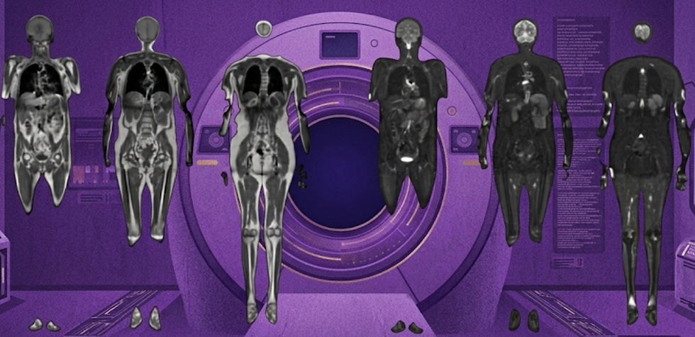
MRIguidance Aim to Bring Synthetic CT Into Routine Care
New clinical data positions MRIguidance to expand beyond pediatrics and surgical planning.

As clinicians seek ways to reduce radiation exposure in patients—and scanner backlogs—image synthesis may offer compelling advantages, such as MRIguidance's BoneMRI which generates a synthetic CT from an MRI. The company has made some inroads into the market, primarily as a method for reducing CT use during surgical planning—particularly in pediatric patients. However, recently MRIguidance has been pushing for non-surgical planning use cases, including with two recent prospective studies on adult populations published in European Radiology and Scientific Reports.
In European Radiology, the multicenter multireader study evaluated BoneMRI’s performance in the cervical spine among 37 trauma participants—with the following key results:
Synthetic CT alone detected 97.3% of fractures—92.7% when facet injuries were included.
Combining synthetic CT with MRI raised detection to 100% for both categories.
The study showed near-perfect agreement on injury classification and morphology, with strong Hounsfield alignment between synthetic and real CT.
In Scientific Reports, a prospective study compared synthetic and conventional CT in 105 lumbar spine cases, reporting:
Synthetic CT performed well for visualizing osteophytes and detecting annulus fibrosus calcifications.
However, it underperformed for osteoporosis assessment, though details on this analysis were limited.
These findings support BoneMRI’s potential beyond pre-surgical workflows, enabling radiation-free bone imaging in more routine spine diagnostics. MRIguidance also garnered a Medicare billing code earlier this year, removing a major barrier to clinical adoption.
Bottom line: MRIguidance is aiming to move beyond surgical planning and pediatric imaging, with new clinical evidence and a Medicare reimbursement code in hand.
DIY MRI Workshop Returns in 2026
Free, hands-on, and open to everyone!
After a successful workshop in May, sign-ups are now open for the next DELTA DIY-MRI event—taking place September 16–18, 2026, at Johns Hopkins University in Baltimore. This free, hands-on workshop invites students, engineers, and researchers to learn the physics and engineering behind magnetic resonance imaging by building their own fully functioning low-field MRI scanner using open-source tools and hardware.
Participants will be guided by a passionate team of mentors dedicated to expanding access to imaging education: Ivan Etoku Oiye, Charlotte Sappo, Sebastian Theilenberg, Sairam Geethanath, Ajay Sharma, Dinil Sasi Sankaralayam, Jonathan Martin, and Zinia Mohanta. Whether you're new to MRI or looking to dive deeper into low-cost scanner development, DELTA DIY-MRI offers a unique opportunity to demystify MRI from the inside out. Learn more and sign up at delta-diy-mri.github.io.
Bottom line: If you want to help build a DIY MRI, sign up here! delta-diy-mri.github.io
Feedback
We’re eager to hear your thoughts as we continue to refine and improve RadAccess. Is there an article you expected to see but didn’t? Have suggestions for making the newsletter even better? Let us know! Reach out via email, LinkedIn, or X—we’d love to hear from you.
References
Fischer, Gregor, et al. "Radiological evaluation and clinical implications of deep learning-and MRI-based synthetic CT for the assessment of cervical spine injuries." European Radiology (2025): 1-13.
Jiang, Ziwei, et al. "Evaluation of MRI-based synthetic CT for lumbar degenerative disease: a comparison with CT." Scientific Reports 15.1 (2025): 20548.
Disclaimer: There are no paid sponsors of this content. The opinions expressed are solely those of the newsletter authors, and do not necessarily reflect those of referenced works or companies.