- Campbell Arnold
- Sep 23, 2025
- 6 min read


"Radiology is at a pivotal inflection point, where smart clinical AI will define scanner performance.”
— Robert Lauritzen, CEO & Co-founder of Cerebriu
Welcome to Radiology Access! your biweekly newsletter on the people, research, and technology transforming global imaging access.
In this issue, we cover:
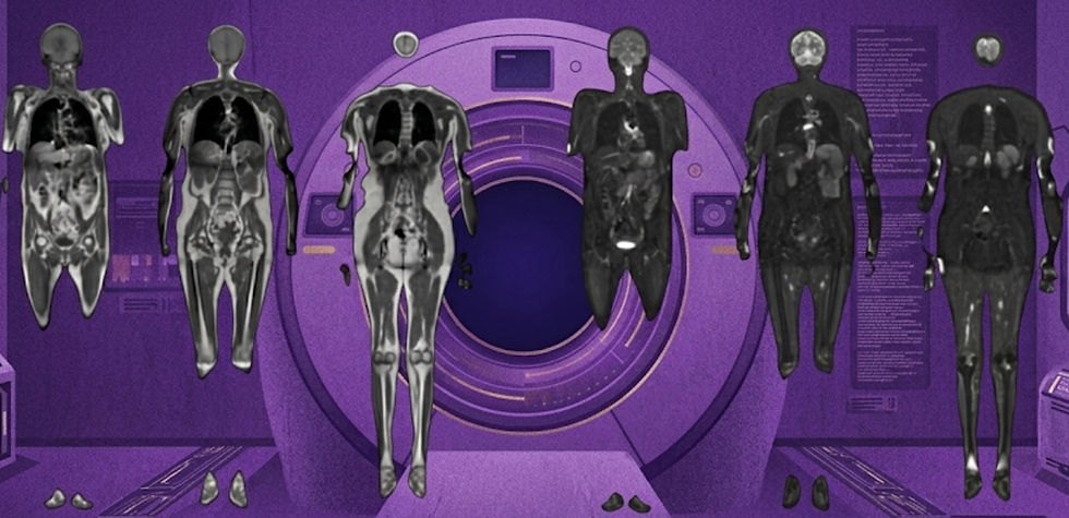
Cerebriu’s Big Week: $11.1M Raise + CE Mark w/ Major OEM
Digital Breast Tomosynthesis Increases Sensitivity w/o More False Positives
Increasing imaging capacity with AI
If you want to stay up-to-date with the latest in Radiology and AI, then don't forget to subscribe!
Cerebriu’s Big Week: $11.1M Raise + CE Mark w/ Major OEM
Could adaptive protocols be coming to a Siemens scanner near you?

In our last issue, we highlighted a European Journal of Radiology study evaluating Cerebriu’s Apollo Smart Protocol, an AI system for real-time MRI protocol adaptation based on detection of infarcts, intracranial hemorrhages, and tumors. Using a 3 or 4 sequence abbreviated protocol, Apollo can analyze images while the patient is still in the scanner and recommend additional sequences when pathologies are detected, like contrast-enhanced imaging for a tumor.
But Cerebriu clearly had more than research in the pipeline. This week, the company announced two major milestones: CE-mark approval for Apollo Smart Protocol and an $11.1M funding round. A particularly interesting aspect of this regulatory clearance is that Cerebriu partnered with Siemens Healthineers to directly embed the algorithm into the OEM’s MRI system. This direct integration is crucial for enabling the kind of fast, in-scanner decisions required for adaptive protocols. The CE-mark clearance gives the technology a pathway into clinical practice in Europe, while the Siemens partnership provides a direct integration channel into MRI systems, paving the way towards real-world adoption.
The $11.1M funding round, led by Denmark’s state-owned Export and Investment Fund, will support commercialization and global scaling. The company is already pursuing FDA clearance and is planning expansions into Asia and the Middle East. For hospitals, the appeal is clear: smart, adaptive MRI protocols could reduce inefficiencies, minimize recalls, and streamline workflows, all at a time when demand for imaging continues to outpace radiologist capacity.
Bottom line: With a CE-mark, Siemens partnership, and $11.1M in new funds, Cerebriu is poised to move its adaptive MRI protocols from research into real-world clinical practice.
Digital Breast Tomosynthesis Increases Sensitivity Without More False Positives
Why you should be using digital breast tomosynthesis for your screening program.

Prior work from the TOmosynthesis plus SYnthesized MAmmography (TOSYMA) trial, published in Lancet Oncology and Radiology, established that digital breast tomosynthesis (DBT) outperforms digital mammography. The landmark trial included nearly 100,000 women across 17 sites in Germany, randomized to DBT with synthetic mammography or standard digital mammography. Initial findings showed a 48% increase in cancer detection rates, with the greatest benefit observed in women with dense breasts.
In their latest Radiology publication, the TOSYMA investigators explored whether this increased sensitivity comes at the cost of higher false positives. They analyzed both false-positive recalls (women recalled for further testing without a cancer diagnosis) and false-positive biopsies (invasive procedures that did not confirm cancer). Results were encouraging: the false-positive recall rate was slightly lower with DBT (40.2 vs. 43.7 per 1,000), and the positive predictive value was higher (17.2% vs. 12.3%). While the false-positive biopsy rate was slightly higher (7.8 vs. 6 per 1,000), the positive predictive value also improved (51.7% vs. 50.6%).
These findings suggest that DBT not only detects more cancers but also improves diagnostic efficiency, reducing unnecessary recalls while maintaining acceptable biopsy trade-offs. For screening programs, this strengthens the case for DBT as a first-line modality, particularly in first round screenings and women with dense breasts. The results also highlight an important broader point: as imaging technology advances, the challenge is not only to improve sensitivity but to do so while minimizing patient burden and maximizing clinical value.
Bottom line: Digital breast tomosynthesis boosts cancer detection without increasing false-positive recalls, making it a strong candidate for first-line breast cancer screening.
Increasing imaging capacity with AI
Some thoughts on why AI is a compelling solution to raising imaging volumes.

At ASFNR in Austin last weekend, I gave a talk on the clinical deployment of AI solutions. In preparing, I took a deep dive into the imaging capacity problem—and I’d like to share some key takeaways.
Two recent JACR studies projected U.S. imaging volumes and radiologist workforce growth through 2055. The message was clear: demand growth will continue to outpace workforce growth, widening the supply-demand gap. Anyone in radiology already feels this shortage, but the question remains—what can we do about it? Broadly, there are three levers to pull:
Decrease imaging volumes
Increase personnel and infrastructure
Improve existing personnel / infrastructure efficiency
The most effective, though least realistic, solution would be to reduce imaging demand. While growth has decelerated from a mid-2000s peak of ~10% annually to today’s ~3–5%, that’s still growth. Population demographics heavily favor continued growth, even if efforts are made to curb unnecessary imaging.
The next lever is expanding the workforce. Radiology residency slots hit a record high in 2025, representing ~3.5% growth in the number of radiologists. However, that doesn’t account for ~1% of radiologists retiring annually and increasing attrition. Accounting for these, supply growth still falls well short of meeting demand increases. Some propose easing restrictions to allow more mid-level providers or outsourcing to international radiologists, but these raise concerns about quality and long-term impact on the profession. Other short-term solutions include asking existing radiologists to simply work more through delayed retirement, fewer vacations, or moonlighting, as suggested in a recent AJR article; However, this is neither sustainable nor appealing. On the infrastructure side, adding scanners and imaging centers to increase our imaging capacity comes with staggering costs.
That brings us to the third lever: making the system we already have more efficient. With ~40,000 US radiologists, a modest 10% productivity gain would equal three full years of new residency cohorts. On the scanner side, the US has ~13,000 MRIs; boosting throughput by 20–25% would be equivalent to deploying 3,000 new scanners, saving roughly $6B in capital costs. Smarter protocols, better scheduling, workflow augmentation, and optimized routing can all play a role here.
This is where AI shines. It’s inherently scalable. Once validated, an algorithm can be deployed across additional scanners or radiologists relatively quickly. It’s also transferable, with the same solution working across sites and institutions. Compared with adding hardware or training new radiologists, it’s lower cost. And importantly, AI integrates into existing IT infrastructure of health care systems. Finally, as a tool, AI is flexible enough to address bottlenecks at multiple points in the imaging workflow.
Bottom line: If imaging demand continues to rise faster than workforce growth, AI may be our best lever to expand capacity, without burning out radiologists or breaking the bank.
Feedback
We’re eager to hear your thoughts as we continue to refine and improve RadAccess. Is there an article you expected to see but didn’t? Have suggestions for making the newsletter even better? Let us know! Reach out via email, LinkedIn, or X—we’d love to hear from you.
References
Sheng, Kaining, et al. "Accuracy of detecting critical findings using abbreviated brain MRI scan protocols as a prerequisite for AI-driven on-the-fly scan protocol adaptation." European Journal of Radiology (2025): 112365.
Weigel, Stefanie, et al. "False-Positive Recall and False-Positive Biopsy Rates in Mammography Screening: A TOSYMA Trial Subanalysis." Radiology 316.3 (2025): e251014.
Heindel, Walter, et al. "Digital breast tomosynthesis plus synthesised mammography versus digital screening mammography for the detection of invasive breast cancer (TOSYMA): a multicentre, open-label, randomised, controlled, superiority trial." The Lancet Oncology 23.5 (2022): 601-611.
Weigel, Stefanie, et al. "Breast density and breast cancer screening with digital breast tomosynthesis: a TOSYMA trial subanalysis." Radiology 306.2 (2022): e221006.
Rawson, James V., Dana Smetherman, and Eric Rubin. "Short-term strategies for augmenting the national radiologist workforce." American Journal of Roentgenology 222.6 (2024): e2430920.
Disclaimer: There are no paid sponsors of this content. The opinions expressed are solely those of the newsletter authors, and do not necessarily reflect those of referenced works or companies.