- Campbell Arnold
- Oct 21, 2025
- 4 min read


"A brute-force approach of simply collecting more data is not an effective path forward.”
— Ilse et al., arXiv 2025
Welcome to Radiology Access! your biweekly newsletter on the people, research, and technology transforming global imaging access.
In this issue, we cover:
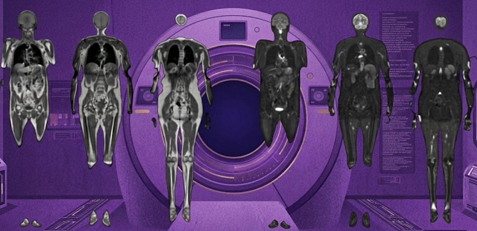
Bigger is Better, but Bespoke is Best: Training Effective Foundation Models
Tomorrow's Scan, Today: Using AI to Predict Future MRIs
Unlocking Quantitative Imaging at Low-Field
If you want to stay up-to-date with the latest in Radiology and AI, then don't forget to subscribe!
Bigger is Better, but Bespoke is Best
Could site-specific pretraining outperform massive open-weight foundation models?

How much data do you really need to train a high-performing foundation model? While most large, open-source vision-language models rely on hundreds of millions of images, datasets in radiology are much smaller, more fragmented, and often siloed for privacy reasons. A recent arXiv preprint systematically explores how radiology foundation model performance scales with dataset size and compares generalized open-weight models to site-specific pretraining.
The authors found that model performance increases predictably with more data, though gains begin to plateau at large scales. Strikingly, they showed that individual centers could outperform open-weight foundation models with as few as 30,000 local samples. This suggests that massive centralized datasets may not be necessary for optimal performance, especially given that local data better reflect the scanners, imaging protocols, and patient populations seen in practice.
The implications for device development are significant. Rather than relying solely on massive generalist models, hospitals could fine-tune smaller, domain-specific foundation models that achieve better accuracy using relatively modest data and compute. This finding dovetails with efforts from companies like Microsoft and HOPPR, which aim to provide initial model weights and infrastructure for institutions to develop in-house algorithms. While this “local foundation model” approach may initially be limited to large health systems and enterprise private practices, it could mark the beginning of a shift toward bespoke, site-optimized AI in radiology.
Bottom line: This study shows that site-specific foundation models can outperform massive open-weight models, highlighting the power of tailored data over sheer scale.
Tomorrow's Scan, Today: Using AI to Predict Future MRIs
Can deep learning predict what your brain will look like in two years?

What if an MRI could show you your future? That’s the vision behind a recent Radiology AI study presenting a deep learning framework that predicts how an individual’s brain might look months or even years later. The work focuses on early childhood development, a period of rapid structural and functional change, and demonstrates the ability to synthesize longitudinal brain MRIs from a single baseline scan.
The team trained their model on longitudinal infant datasets spanning 2 weeks to 24 months, enabling it to generate anatomically realistic MRIs that reflect expected developmental trajectories. Using a dual-stage U-Net architecture with time-conditioning, the network can simulate both forward and backward changes, reconstructing how a child’s brain may have looked in the past or will look in the future. The synthesized future scans closely resembled real follow-up images, supported by strong quantitative results (SSIM = 0.87). The scans also preserve subject-specific anatomy and capturing population-level developmental trends, such as changes in cortical thickness, white matter maturation, and regional volumes.
Beyond its technical achievement, the study opens a new opportunities for longitudinal neuroimaging studies and offers the potential to predict an individual's development. It will be fascinating to see how such algorithms perform in children with atypical developmental trajectories.
Bottom line: This study presents an algorithm that uses time conditioning to predict future and past MRI scans from a present study, which could aid in predicting pediatric brain development.
Unlocking Quantitative Imaging at Low-Field
How researchers built a 10 minute T₁ mapping protocol on Hyperfine’s Swoop.

Quantitative T₁ mapping has become an important biomarker, offering insights into healthy myelination in children and disease processes, like demyelination in multiple sclerosis and structural changes in Alzheimer’s. However, achieving robust T₁ maps at low-field strengths is challenging due to lower SNR, long scan times, and electromagnetic interference in unshielded environments.
In a new multicenter study published in Imaging Neuroscience, researchers evaluated a 10.8-minute inversion-recovery T₁ mapping protocol on Hyperfine’s portable 64 mT MRI across six international sites. Using a phantom developed by CaliberMRI, the team determined inter-site differences in T₁ measurements were under 3%. They also performed 60 in vivo scans, establishing T₁ values (approximately 290 ± 6 ms for white matter and 332 ± 8 ms for cortex), recording low inter-site variability, and also showing longitudinal stability.
These results demonstrate that T₁ mapping at 64 mT can be achieved with high repeatability and reproducibility. Crucially, this unlocks additional quantitative capabilities on these low field systems that complements recent qualitative gains in image quality.
Bottom line: This multicenter study proves that reliable, quantitative T₁ mapping is achievable on portable 64 mT MRI systems in 10 minutes.
Feedback
We’re eager to hear your thoughts as we continue to refine and improve RadAccess. Is there an article you expected to see but didn’t? Have suggestions for making the newsletter even better? Let us know! Reach out via email, LinkedIn, or X—we’d love to hear from you.
References
Ilse, Maximilian, et al. "Data Scaling Laws for Radiology Foundation Models." arXiv preprint arXiv:2509.12818 (2025).
Fang, Yu, et al. "A Deep Learning Framework for Synthesizing Longitudinal Infant Brain MRI during Early Development." Radiology: Artificial Intelligence (2025): e240708.
Lena, Beatrice, et al. "Repeatability and reproducibility of rapid T1 mapping of brain tissues at 64 mT: a multicentre study." Imaging Neuroscience (2025).
Disclaimer: There are no paid sponsors of this content. The opinions expressed are solely those of the newsletter authors, and do not necessarily reflect those of referenced works or companies.