- Campbell Arnold
- May 6, 2025
- 7 min read


“We believe that in 10 years low field MRI will be as ubiquitous as routine blood sampling. It will be used to screen for cancer, in constrained settings like the operation room, [and] to build large quantitative models for personalized medicine.”
— Evan Kervella, Chipiron Co-founder & CEO
Welcome to Radiology Access! your biweekly newsletter on the people, research, and technology transforming global imaging access.
You might notice a few changes in this issue—thanks to a thoughtful reader, Anna Menyhart-Borroni, who reached out with some great suggestions! I always love hearing from people who read the newsletter, and I’m incredibly grateful for the feedback. Love it? Hate it? Let me know—just shoot me an email!
In this issue, we cover:
Want to stay up-to-date with the latest in Radiology and AI? Then don't forget to subscribe!
Chipiron Raises $17M to Reinvent MRI for the Frontlines
And how their technology differs from other low-field scanners.

MRI is one of the most powerful tools in modern medicine, but despite its clinical value, it remains out of reach for many. High costs, massive footprints, and infrastructure requirements have historically confined scanners to hospitals or specialized imaging centers, often far from where care is urgently needed. Chipiron, a Paris-based startup, just announced a $17 million Series A funding round to build a lightweight and affordable ultra-low-field MRI designed for point-of-care use.
Low-field MRI isn’t a new idea — in fact the first ever MRI was 50 mT, and in recent years low-field strengths have received renewed commercial interest. What sets Chipiron apart is how low they can go. While most low-field systems operate in the 50–100 mT range using heavier permanent magnets, Chipiron is going to true ultra-low-field strengths (under 10 mT). This enables the use of resistive magnets which are lightweight, open, and allow device designs to adapt to constrained environments like ICUs and operating rooms.
“We believe the reason low-field MRI hasn't found product-market fit yet lies in product design.” said Chipiron CEO Evan Kervella. “Going to ultra-low fields [can accommodate] any form factor and any constrained setting. It unlocks full body imaging.”
This design choice opens the door to new use cases and places where conventional systems simply can’t go — potentially expanding MRI use for routine screenings, intraoperative guidance, and remote care.
With the new infusion of cash, Chipiron plans to:
Begin EU clinical deployment in 2025
Collect in-vivo studies and select an initial use case
Grow their technical, product, and regulatory teams
Begin the FDA filing process in 2026
With a bold design philosophy and now a solid runway, Chipiron joins a growing wave of startups trying to make MRI as accessible as radiography.
Bottom line: Chipiron is betting on a lightweight, flexible ultra-low-field design to break MRIs out of the hospital basement — now they have $17M to help them do it.
LowGAN: Generating High-Quality Images from Low-Field MRI
Using AI to bridge the field strength gap & expand access to diagnostic imaging.

This week, I'm excited to be able to cover my own work, recently published in Radiology! This project is especially meaningful to me, as it marks the end of my PhD era. I started this work near the end of my PhD, and I'm incredibly grateful to the team at UPenn for helping see it through to completion. It also combines two of my favorite research topics: low-cost imaging and AI image enhancement.
In the paper, we developed an image enhancement algorithm called LowGAN, designed to process scans collected on the Hyperfine portable MRI. In the study, we:
Collected paired 64mT and 3T scans of MS patients.
Trained a GAN to synthesize 3T-like images from 64mT sequences.
Quantified improvements in image quality, volume measures, and lesion measures.
Overall, we found:
Synthesized images were quantitatively more similar to high-field images.
Brain volume measurements showed improved accuracy.
White matter lesions became more conspicuous — though lesions not visible in the original 64mT scans were not resolved.
To me, this work illustrates both the power and the limitations of AI for improving image quality and highlights the potential for portable MRI systems to broaden access to high-quality imaging.
Bottom line: LowGAN shows how AI can enhance low-field MR image quality, improving clinical utility and helping expand access to advanced neuroimaging.
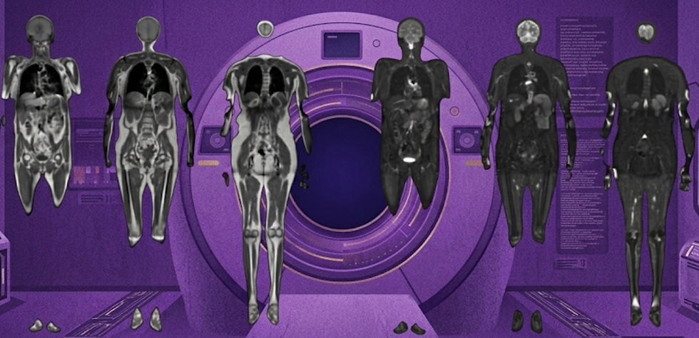
Function Health Acquires Ezra, Offers $499 Full-Body MRI Scans
And what consolidation means for the preventive healthcare care space.

In a move that took much of the radiology industry by surprise, Function Health—a startup best known for its blood diagnostics platform—announced yesterday that it has acquired Ezra, a company behind full-body MRI scans aimed at early detection.
The acquisition marks a major step toward integrated preventive screening, combining both imaging- and blood-based diagnostics under one platform. But just as notable as the acquisition is the pricing strategy: Function is coming in aggressively, signaling a race to the bottom in cost.
A quick look at the numbers:
Prenuvo, Ezra’s main competitor, offers a 45-minute, head-to-ankle scan for$2,499 and a 20-minute torso scan for $999.
Ezra had been offering a 30-minute, 4-region scan (brain, neck, abdomen, pelvis) for $1,495 with a spine add on for $2395.
Function has now slashed the price to $499 for a 22-minute scan—presumably covering 3–4 regions—dramatically undercutting the competition.
The preventative care space is heating up. Earlier this year, Prenuvo raised $120M to expand their offerings and geographical coverage. Function and Ezra have together raised $97M, with plans to raise an additional $200M. The appetite for proactive health services appears strong: Personally, I’ve spoken to consumers who’ve purchased these products—all of whom seem generally satisfied. I’ve also spoken to radiologists reading these scans—many reporting highly competitive compensation.
Still, a dose of realism: at $499 a scan, it takes a lot of volume to justify a ~$300M investment! Whether this new price point can support a sustainable business—or shift consumer behavior on a mass scale—remains to be seen. Function is likely betting that affordable MRI will act as a gateway, attracting new users who will stick around for biannual blood testing and ongoing health subscriptions. One thing is clear: AI-driven reductions in scan time are rapidly transforming full-body MRI from a luxury product into a more accessible tool for preventive care.
Bottom line: With its acquisition of Ezra and price cut to $499 for full-body scans, Function Health is betting big on making preventive care mainstream, and that consumers will stick around as subscribers.
Health Tech Investment Act Introduced in Congress
And what that means for Radiology AI.

Recent developments in the US are paving the way for improved reimbursement pathways for AI-driven radiology tools, potentially accelerating their clinical adoption. As it stands, there is no guarantee that after your device is approved that medicare will provide reimbursement. In fact, the early reimbursement is a patchwork of temporary codes that may or may not lead to established, long term reimbursement.
A recent bipartisan bill, the Health Tech Investment Act (S.1399), has been introduced in Congress to establish a more predictable reimbursement framework for FDA-approved AI-enabled medical devices. The legislation proposed to streamline device approval to reimbursement pathway by:
Assigning all FDA-cleared products a Category III New Technology Ambulatory Payment Classification, providing a structured reimbursement framework.
Monitoring codes for five years to enable cost data collection before assigning a permanent payment code.
In the meantime, radiology AI companies aren’t waiting around. May have braved the system to obtain codes are start establishing themselves as reimbursable products:
Last month MRIguidance's BoneMRI, which generates CT-like images using MRI scans, received a Medicare billing G-code (G0566).
Last year Icometrix established broad CPT Category III codes (0865T and 0866T) covering lesion detection and quantitative analysis in the brain.
These advancements reflect a broader trend toward integrating AI into medical imaging, with legislative support and specific company achievements indicating a move toward more accessible and reimbursable AI-driven diagnostic tools. This is particularly crucial for companies that have AI products that improve the quality of care, but have struggled to quantify how that impacts a healthcare organization’s bottom line.
Bottom line: A new bipartisan bill signals that radiology AI is inching closer to a sustainable reimbursement model—crucial for long-term adoption, especially for companies that can’t clearly demonstrate ROI.
Resource Highlight: Pranav Rajpurkar
For this week's resource highlight, I’m recommending you follow Pranav Rajpurkar (LinkedIn, X). Pranav is an Assistant Professor of Radiology at Harvard and co-founder of a2z Radiology AI.
In the last 3 weeks alone, he and his associates have released 3 massive datasets:
ReXGradient-160K: A Large-Scale Multi-Institutional Chest X-Ray Dataset
ReXErr-v1: Clinically Meaningful Chest X-Ray Report Errors Derived from MIMIC-CXR
Collab-CXR: a unique resource to study human-AI collaboration in chest X-ray interpretation.
If you want to learn more about Pranav, I highly recommend you listen to his episodes on the Radiology AI podcast and the NEJM AI Grand Rounds podcast.
Feedback
We’re eager to hear your thoughts as we continue to refine and improve RadAccess. Is there an article you expected to see but didn’t? Have suggestions for making the newsletter even better? Let us know! Reach out via email, LinkedIn, or X—we’d love to hear from you.
References
Lucas, Alfredo, et al. "Multisequence 3-T image synthesis from 64-mT low-field-strength MRI using generative adversarial networks in multiple sclerosis." Radiology 315.1 (2025): e233529.
Ashley Capoot. “Function Health buys Ezra, launches full-body scan for a third of the price.” CNBC 2025.
Onac, Laura, et al. "An image-domain deep-learning denoising technique for accelerated parallel brain MRI: prospective clinical evaluation." Radiology Advances 1.3 (2024): umae022.
https://theimagingwire.com/2025/04/13/payment-path-for-medical-ai/
https://mriguidance.com/bonemri-receives-medicare-billing-code/
Moehring, Alex, et al. "A Dataset for Understanding Radiologist-Artificial Intelligence Collaboration." Scientific Data 12.1 (2025): 739.
Disclaimer: There are no paid sponsors of this content. The opinions expressed are solely those of the newsletter authors, and do not necessarily reflect those of referenced works or companies.