- Campbell Arnold
- Apr 28
- 7 min read

“Holding a 510(k) clearance may not reflect that a manufacturer is proficient in, or even has experience with, the processes used in the development of the cleared device.”
— Ellen J. Flannery, Director, Office of Policy, FDA
Welcome to Radiology Access! Your biweekly newsletter on the people, research, and technology transforming global imaging access.
In this issue, we cover:
Super-resolution reconstruction sharpens stenosis classification in CCTA
The Rise of Regulatory Arbitrage in Medical Imaging AI
Portable MRI for Lowering Barriers to Global Dementia Care
If you want to stay up-to-date with the latest in Radiology and AI, then don't forget to subscribe!
How new hardware can improve the quality of our existing scanner fleet
How Canon's new super-resolution reconstruction sharpens CCTA stenosis classification on traditional scanners, using newer scanner data.
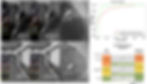
Coronary CT angiography has become a frontline tool for evaluating coronary artery disease, but its limited spatial resolution can blur lesion margins just enough to push a borderline stenosis into the wrong category, altering CAD-RADS classification, downstream referrals, and ultimately patient management. Newer photon-counting CT (PCCT) systems have shown superior performance for stenosis assessment, but their cost remains a major barrier to broad clinical adoption. But what if rather than waiting for an expensive scanner replacement, we could leverage data from high resolution systems to extract more diagnostic value during reconstruction on our conventional systems?
In a new Radiology study, the authors evaluated Canon’s super-resolution deep learning reconstruction algorithm, trained on 0.25 mm high-resolution data and applied to conventional CCTA acquisitions. In a prospective, blinded, multi-center study spanning 10 sites in China, the model significantly improved stenosis assessment compared with hybrid iterative reconstruction, using invasive coronary angiography as the reference standard. The gains were not merely visual, they were clinically meaningful. Lesion-level AUC improved from 0.90 to 0.97, and CAD-RADS classification changed in 20% of patients, directly affecting diagnostic categorization and downstream management.
What makes this work especially compelling is that it demonstrates a more immediately actionable method for improving diagnostic quality. Software can deliver clinically meaningful image quality gains without requiring new hardware. That matters because history has shown that high-end imaging systems, such as PCCT, 7T MRI, and total-body PET, often take years to move beyond flagship centers. Super-resolution reconstruction offers a faster path for those performance gains to reach routine care, allowing advances in scanner design to influence clinical workflows more broadly long before full hardware adoption catches up.
If super-resolution reconstruction can reliably reclassify disease severity, then it is more than just a better way to turn sinograms into images. It is diagnostic quality improvement embedded directly into reconstruction. The next step is proving that sharper images lead to better decisions, fewer unnecessary procedures, and better outcomes at scale.
Bottom line: Super-resolution reconstruction shows that some of the diagnostic benefits of better hardware can be transferred to existing scanners through smarter reconstruction.
The Rise of Regulatory Arbitrage in Medical Imaging AI
How regulatory bodies are quietly reshaping where and how radiology AI gets built.

Several key regulatory decisions this month are making it increasingly clear that the next phase of medical imaging AI will be shaped less by model performance alone, and more by who can navigate regulation, reimbursement, and scale first.
In South Korea, the Ministry of Food and Drug Safety (MFDS) cleared Soombit.ai’s AIRead-CXR, the world’s first generative AI device for image interpretation. AIRead-CXR is a chest X-ray vision-language model that generates preliminary radiology reports for an eye-popping 57 clinical findings. To put that in context, the largest FDA clearance to date (Aidoc’s CARE) spans 14 indications. The approval of Soombit.ai’s AIRead-CXR is therefore more than a product milestone, it is a clear signal that generative AI in radiology is moving from demos into regulated clinical workflows. Importantly, Korea didn’t simply react to generative AI devices, it preemptively published dedicated guidance and a framework to support pivotal, multicenter clinical trials and a regulatory pathway.
While the FDA has also become more flexible in recent years, it is still signaling a more cautious stance on generative AI. Earlier this month, the agency rejected Harrison.ai’s petition to streamline clearance for multi-model AI developers, declining to create a pathway where additional algorithms could be deployed under post-market oversight rather than requiring new regulatory submissions for each model. For companies building multi-disease AI portfolios, the message was clear: the FDA is still fundamentally regulating AI one model at a time. That approach preserves a slower, more conservative framework at a time when model development itself is accelerating rapidly. However, CMS and the FDA did announce a new streamlined pathway to align breakthrough device designation and reimbursement, which addresses a different but equally critical bottleneck.
The rapidly changing regulatory landscape may offer a new form of regulatory arbitrage in medical AI. The next winners in imaging AI may not be the companies with the best-performing models, but those best aligned with the regulatory environments they choose to enter. One reason companies like RadAI have been able to iterate quickly is that they’ve operated in a relatively unconstrained regulatory gray area in the US, allowing for rapid deployment and refinement. That window could narrow, but other opportunities are opening up. Increasingly, strategy may be defined less by market size alone, and more by where regulatory pathways are fastest, clearest, and most advantageous. The frontier is no longer just model performance, it is regulatory design as competitive advantage.
Bottom line: The rapidly changing and increasingly divergent regulatory and reimbursement landscape across countries may offer companies unique opportunities for faster, broader clearances.
Portable MRI for Lowering Barriers to Global Dementia Care
How low-field MRI could expand access and reshape Alzheimer’s neuroimaging.
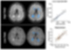
Alzheimer's disease and related dementias are rising worldwide, but the infrastructure needed to diagnose and monitor them remains profoundly uneven. Over the next 25 years, dementia prevalence is projected to grow fastest in low- and middle-income countries (roughly 58–71% versus 30–42% in high-income regions), yet access to MRI remains concentrated in urban areas and high-income countries, where scanner density can be more than 100-fold higher per capita. The result is a widening mismatch between where neurodegenerative disease burden is growing fastest and where advanced imaging is actually available. A recent Nature Reviews Neurology article argues that portable low-field MRI could help close that gap, lowering the barriers to imaging access in dementia care while opening new opportunities for diagnosis, monitoring, and research.
Low-field MRI is not simply a cheaper scanner, it is a different deployment model for neuroimaging. Compared with conventional systems, portable low-field platforms are less expensive to install and operate, require less infrastructure, and can be deployed closer to the patients who need them. That creates several immediate opportunities in dementia care:
Expanded imaging access: Lower-cost systems can increase scanner availability beyond major urban centers, reducing travel burden and long wait times.
More accessible longitudinal monitoring: Broader deployment could make repeat MRI more feasible for tracking disease progression and treatment response.
More representative research: Expanding MRI access beyond major academic centers could improve inclusion in neurodegeneration studies, particularly in regions carrying the greatest disease burden.
Reduced workforce constraints: Many low-field systems are easier to operate than conventional MRI, lowering training barriers and expanding who can acquire clinically useful scans.
The technical tradeoff, of course, is lower signal and lower native resolution, but that gap is narrowing quickly. Advances in reconstruction, denoising, super-resolution, and segmentation have made low-field systems increasingly capable of capturing clinically meaningful neurodegenerative biomarkers, including regional atrophy and white matter hyperintensities. That progress is already visible in practice: Hyperfine’s portable MRI platform has demonstrated how much image quality gains can be recovered through deep learning based reconstruction and post-processing.
What makes portable MRI especially compelling in dementia care is not that it will replace conventional high-field systems (it won’t), but that it can extend how and where neuroimaging is used For Alzheimer disease several applications stand out:
Brain volumetrics: Measuring regional volume changes, such as hippocampal atrophy, can help distinguish healthy aging from neurodegenerative disease and support earlier diagnosis.
White matter hyperintensities: Tracking white matter lesion burden has implications for cognitive decline in Alzheimer disease and may also inform care in related neurologic conditions, including vascular disease and multiple sclerosis.
DWI measurement: Diffusion-weighted imaging may help detect white matter microstructural and vascular injury in regions affected by dementia pathology, though this application still requires further validation at low field.
ARIA monitoring: Patients receiving anti-amyloid therapies require serial imaging to monitor amyloid-related imaging abnormalities (ARIA), and low-field MRI has shown early promise for detecting at least some of these treatment-related complications.
The long-term opportunity is bigger than portability alone. Low-field MRI offers a chance to rethink how neuroimaging is distributed globally and to align imaging access more closely with where dementia care is needed most.
Bottom line: Portable low-field MRI offers a practical path to expanding dementia imaging access globally by bringing clinically useful neuroimaging closer to the patients who need it most.
Feedback
We’re eager to hear your thoughts as we continue to refine and improve RadAccess. Is there an article you expected to see but didn’t? Have suggestions for making the newsletter even better? Let us know! Reach out via email, LinkedIn, or X—we’d love to hear from you.
References
Zou, Limiao, et al. "Super-Resolution Deep Learning Reconstruction for Coronary CT Angiography: Coronary Stenosis Assessment and CAD-RADS Reclassification." Radiology 318.2 (2026): e252163.
Hwang, Eui Jin, et al. "Clinical validation of a generative artificial intelligence model for chest radiograph reporting: a multicohort study." Radiology 316.3 (2025): e250568.
Sorby-Adams, Annabel J., et al. "Portable, low-field magnetic resonance imaging for evaluation of Alzheimer’s disease." Nature Communications 15.1 (2024): 10488.
Disclaimer: There are no paid sponsors of this content. The opinions expressed are solely those of the newsletter authors, and do not necessarily reflect those of referenced works or companies.