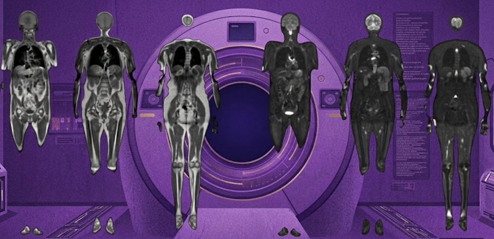
“AI in radiology is moving beyond image interpretation.”
— SIIM 2026 recap by Dr. Amine Korchi, radiologist and industry thought leader
Welcome to Radiology Access! Your biweekly newsletter on the people, research, and technology transforming global imaging access.
In this issue we cover:
Subtle Medical Secures $33M in New Funding, Launches PET and CT Products
MRIxFields 2026: Can AI Bridge the Gap Between Image Quality and Accessibility?
SIIM 2026: AI Is Moving Beyond Image Interpretation
If you want to stay up-to-date with the latest in Radiology and AI, then don't forget to subscribe!
Subtle Medical Secures $33M in New Funding, Launches New PET and CT Products
What do these new funds and product launches mean for the company?

Fair warning, what you're about to read is not objective journalism. It's the enthusiastic perspective of a company insider who has been eagerly waiting to talk about these announcements.
Over the past few weeks, Subtle Medical has unveiled a series of developments that paint a clear picture of where the company is headed, beyond MRI and toward becoming a multimodality imaging platform. New FDA clearances, new funding, and new leadership all point toward the goal of building a vendor-agnostic AI platform that helps health systems get more value from their existing imaging infrastructure.
The momentum began with the FDA clearance of SubtleHD™(PET), announced just ahead of SNMMI in Los Angeles. As PET demand continues to rise, fueled by theranostics and an expanding radiotracer portfolio, imaging providers face increasing pressure to expand capacity. SubtleHD™(PET) addresses that challenge by enabling faster PET imaging while improving image quality. Notably, the broad clearance covers all FDA-approved radiotracers and supports both PET/CT and PET/MR systems, positioning the software to scale alongside the rapidly growing molecular imaging market.
Just days later came another major announcement, Subtle Medical closed a $33 million series C led by Morgan Stanley Expansion Capital. The round also included participation from Shinhan Venture Investment and strong follow-on investments from existing investors, including Fusion Fund, EnvisionX, BRV Capital Management, and Samsung Ventures.
Alongside the financing, the company announced the appointment of industry veteran Ohad Arazi as Chief Executive Officer. “What drew me to Subtle is that this isn't a point solution—it's becoming the AI layer where health systems run their imaging,” Arazi said.
That statement became even more meaningful a week later when Subtle announced FDA clearance for SubtleHD™(CT), the company's first CT product. With the addition of CT, Subtle now offers FDA-cleared image enhancement solutions across three major modalities, MRI, PET, and CT. Rather than requiring health systems to manage separate software products across scanners, vendors, and modalities, Subtle has positioned itself as a single enterprise solution capable of serving an organization's entire imaging fleet.
Taken together, these announcements signal a broader strategic shift. Subtle appears increasingly focused on becoming the centralized AI platform for multimodality image enhancement and workflow optimization. The vision is not simply to improve one exam type, but to create a consistent AI infrastructure layer that operates across vendors, modalities, and clinical environments. Despite the growing scope, Subtle co-founder and Chief Scientific Officer Enhao Gong emphasized that the company remains committed to its original mission of "making advanced imaging faster, safer, and more accessible."
For those of us inside the company, it's been exciting to watch these pieces come together. More importantly, it's exciting to imagine what comes next.
Bottom line: Subtle Medical’s recent $33M series C, leadership expansion, and CT product expansion signal a transition from a collection of products to a unified AI acquisition platform.
MRIxFields 2026: Can AI Bridge the Gap Between Image Quality and Accessibility?
Could deep learning help bring both low-field and ultra-high-field MRI innovations into clinical practice?

I've watched firsthand as low-field MRI image quality has been transformed by deep learning reconstruction and image enhancement. One of my earliest scans on a Hyperfine system in 2019 barely resembles the images being produced today after the introduction of AI-powered reconstruction and post-processing. The improvement has been remarkable.
At the opposite end of the spectrum, MRI manufacturers have continued pushing toward ever-higher field strengths. In recent years there has been a lot of 7T scanner research, the introduction of the first 5T system in 2024, and the publication of the first human brain images acquired on an 11.7T MRI scanner. The anatomical detail produced by these systems is extraordinary, but so are the costs, which has stifled clinical adoption.
Despite innovation at both extremes, the vast majority of MRIs worldwide are still 1.5T and 3T systems. Low-field MRI has struggled to establish a large enough clinical niche despite its lower cost and portability. Meanwhile, ultra-high-field MRI offers exceptional image quality but remains confined largely to research centers.
Yet interest from the imaging research community has never been higher. Low-field MRI continues to gain momentum, with more than 35 ISMRM abstracts this year involving Hyperfine systems alone. At the same time, researchers continue pushing the boundaries of ultra-high-resolution imaging using increasingly powerful scanners.
This raises an interesting question, what if we could combine the strengths of both approaches?
Rather than waiting for widespread deployment of expensive ultra-high-field systems, AI could potentially learn from them. By leveraging high-quality, ultra-high-field data, researchers may be able to develop image enhancement and synthesis algorithms that improve the quality of lower-cost scanners while preserving their accessibility advantages.
That's the idea behind MRIxFields 2026, a new MICCAI challenge built around what may be the largest publicly available cross-field-strength MRI dataset to date. The dataset includes:
Five field strengths: 0.1T, 1.5T, 3T, 5T, and 7T
Three sequence types: T1-weighted, T2-weighted, and T2-FLAIR
1,900+ unpaired scans
600 paired scans (40 participants × 5 field strengths × 3 sequences)
The competition is currently accepting submissions through September 10th and offers $21,000 in total prizes. Participants will compete across three challenges:
Ultra-high-field (7T) MRI synthesis from arbitrary field strengths
Higher-field MRI generation from ultra-low-field (0.1T) scans
Controllable field-to-field MRI synthesis using a conditional model
While AI won't magically turn a 0.1T scanner into a 7T scanner, competitions like MRIxFields represent an important step toward understanding how much information can be recovered, enhanced, or translated across field strengths. If successful, these approaches could help democratize advances that would otherwise remain locked inside expensive research systems for years or even decades.
Bottom line: MRIxFields 2026 is more than a competition, it's a test of whether AI can help bridge the longstanding tradeoff between MRI accessibility and image quality.
SIIM 2026: AI Is Moving Beyond Image Interpretation
Could the best opportunity to improve imaging efficiency be outside of the reading room?

I finally made it to SIIM this year, and after hearing about it for years, I can confidently say the conference lived up to its reputation. Compared to other more clinical conferences, one theme stood out to me: SIIM 2026 was about everything except image interpretation.
For the better part of a decade, radiology AI has been dominated by algorithms designed to detect findings, segment anatomy, and assist with diagnosis. Those applications are still important, but they were no longer the center of attention in Pittsburgh.
Instead, the spotlight was on workflow efficiency. Companies showcased tools for patient communication, scheduling, protocoling, operational analytics, workflow optimization, and AI-native PACS experiences. Many of these applications can reach the market faster than traditional diagnostic AI because they face fewer regulatory hurdles. Startup founders have undoubtedly taken note of the success of companies like Rad AI, which achieved rapid adoption through workflow-focused, class I products which don’t require FDA submissions, such as automated impression generation. More broadly, it reflects a growing realization that some of healthcare's biggest inefficiencies exist outside the imaging itself.
A second trend that was impossible to ignore was the growing prominence of vision-language models (VLMs). Nearly every vendor seemed to be demonstrating either a reporting copilot, a VLM integration, or a broader foundation model strategy. The conversation is rapidly shifting from "Can generalist VLMs compete with task-specific models?" to "How can VLMs improve radiologist workflows?" Unsurprisingly, the most common use case on the showroom floor was report generation, with foundation models producing draft reports that radiologists could then review, edit, and approve.
Taken together, these trends suggest that the next phase of radiology AI may be defined less by direct image interpretation and more by workflow orchestration. The winners may not be the companies with the best standalone algorithms, but those that can seamlessly integrate AI throughout the imaging enterprise.
Bottom line: Radiology AI is increasingly moving beyond image interpretation and into workflow optimization. At SIIM 2026, it felt like the industry was no longer just asking how AI can help radiologists, but rather how it could help run a radiology department.
Feedback
We’re eager to hear your thoughts as we continue to refine and improve RadAccess. Is there an article you expected to see but didn’t? Have suggestions for making the newsletter even better? Let us know! Reach out via email, LinkedIn, or X—we’d love to hear from you.
References
Disclaimer: There are no paid sponsors of this content. The opinions expressed are solely those of the newsletter authors, and do not necessarily reflect those of referenced works or companies.